
Laboratory of
Ocular Biomechanics
University of Pittsburgh
Current Work
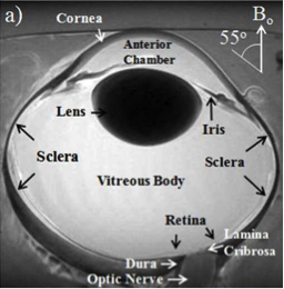
| An axon-centric approach to intraocular-pressure (IOP)-induced mechanical insult | Measuring changes in the optic nerve head during acute and chronic increases in pressure | Using ultrafast lasers for visualizing and modifying collagen microarchitecture and mechanics |
 |
 |
 |
This approach allowed us to discern longitudinal and transverse mechanical insults from an axon perspective, suggesting different axon damage mechanisms. |
We developed a high-resolution digital volume correlation (DVC) method to measure in vivo strains on monkey eyes from optical coherence tomography images. We used this technique to measure the effects of IOP in eyes when they were healthy and at glaucoma onset. The results showed that the eyes at onset had higher strains than those at baseline, suggesting that LC becomes more compliant in early glaucoma. |
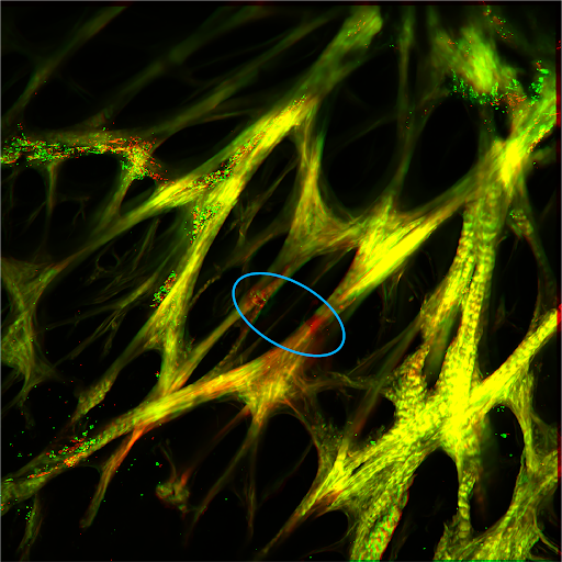
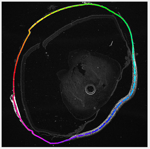
We propose that this will allow improving the mechanical environment of the lamina cribrosa independently of the intraocular pressure. This SHG image shows sheep lamina cribrosa before (red) and after (green) photoablation of a beam (blue oval). |
| A workflow for 2D/3D nuclei segmentation and morphological analysis | A workflow for lamina cribrosa beam segmentation and morphological analysis | A tool for blood flow visualization |
 |
 |
 |
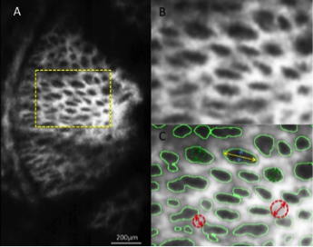
We developed a workflow for 2D/3D nuclei segmentation in ocular tissues and morphological analysis. |
We developed a workflow for segmenting lamina cribrosa beams and analyzing their shape and microstructure at baseline and under load. |
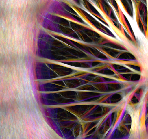
We developed a tool to visualize vasculature structure and animate blood flow within the vascular network. Shown here is the vascular network of a monkey lamina cribrosa region colored by blood flow. |
| A workflow for crimp measurement across corneoscleral shell | Uncovering and quantifying optic nerve head astrocyte morphology | Investigating optic nerve head astrocyte mechanics, in situ |
 |
 |
 |
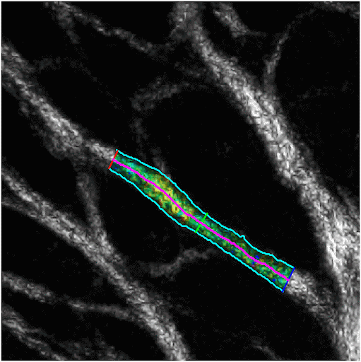
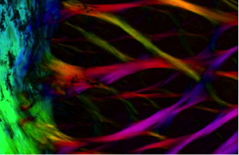
We developed the tools from tissue processing, imaging and analysis to quantify collagen fiber crimp properties over the whole corneascleral shell. |
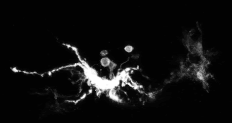
We adapted Multicolor DiOlistic labeling (MuDi) to reveal and quantify in situ optic nerve head astrocyte structure. We are now using this tool to study the astrocytes of several species. |
We reveal the deformations of individual astrocytes due to increased intraocular pressure. Astrocytes have long been suspected to be affected by elevated pressure. Their response to pressure is then believed to contribute to neural tissue damage and glaucoma. |
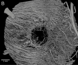
| High-resolution full-depth visualization of the intact lamina cribrosa | Visualizing lamina cribrosa microvasculature | Direct fiber model of the Optic nerve head (ONH) |
 |
 |
 |
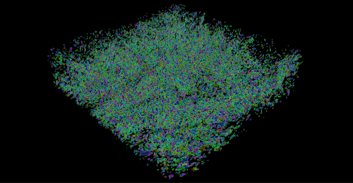
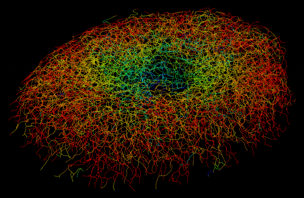
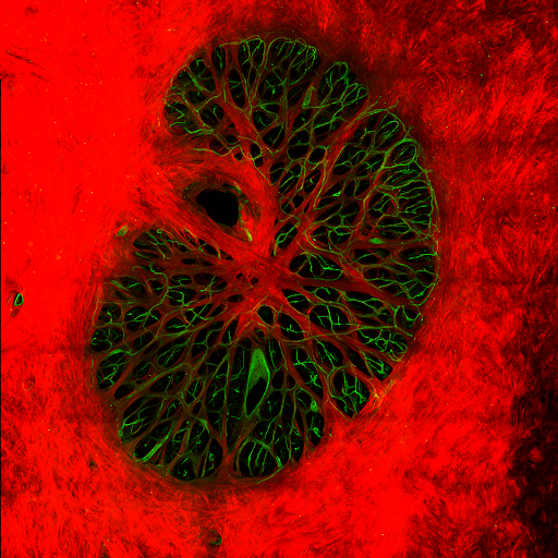
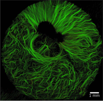
We visualize microstructure of the lamina cribrosa in 3D through tissue clearing and multiphoton microscopy. The beautiful architecture of the collagen that protects the neural tissues becomes visible. We couple this with computational models to understand how this complex structure works. |
We map the interconnected network of microvasculature in the lamina cribrosa. This information is then fed to computational models of blood flow and oxygenation to understand the normal and pathologic optic nerve head. |
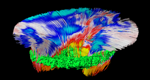
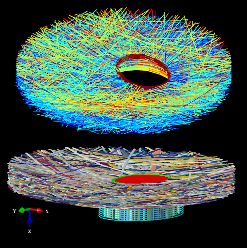
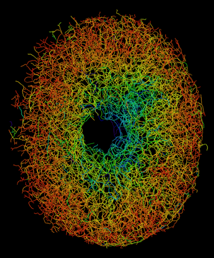
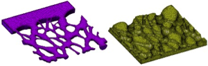
We developed direct fiber models that incorporate the 3D collagen structure and fiber interactions to study ONH biomechanics. These model outperform conventional continuum models by accurately resolving 3D tissue level strain patterns, and preserving long-distance strain transmission along fiber bundles. |
| Hemodynamics of lamina cribrosa vasculature | Structure-function relationship in soft tissues | |
 |
 |
|
We reconstructed the vascular network within the lamina cribrosa region and utilized computational modeling to simulate hemodynamics and oxygenation. This approach allowed us to predict both blood flow and oxygen supply within the lamina cribrosa while assessing the effects of intraocular pressure and remodeling. |
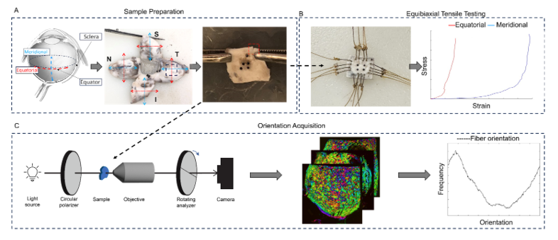
Using polarized-light microscopy and biaxial tensile testing we characterized mechanical and structural anisotropies of the equatorial sclera. Surprisingly, our results show that the structural and mechanical anisotropies do not always coincide. The origin for this difference remains unclear. |
Past Work
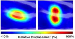
| Direct measurement of 3D deformations of the eye | Effects of aging | Interplay between intraocular and intracranial pressures |
 |
 |
 |
We have developed experimental and computational techniques to measure the acute and chronic displacements and deformations of the eye. |
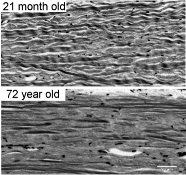
Age is one of the main risk factors for many diseases. We work to understand how aging affects the eye at the microstructural level. |
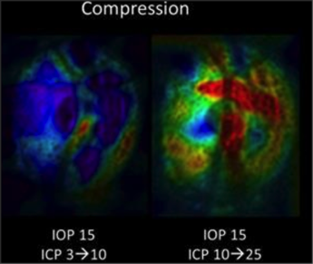
We have developed computational and experimental techniques to study the independent and interacting effects of intraocular and intracranial pressures on the optic nerve head. In collaboration with Gadi Wollstein (NYU) and Matthew Smith (Pitt). |
| Role of eye biomechanics on drusen | Novel imaging tools to study soft tissue collagen | Novel techniques for glaucoma diagnosis |
 |
 |
 |
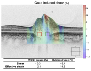
In collaboration with Patrick Sibony (Stonybrook), we are exploring the potential role of biomechanics on the progression of drusen. |
We have developed tools that enable measuring the full 3D orientation of collagen fibers. |
In collaboration with the Glaucoma imaging group (NYU), we are working to develop novel techniques for the diagnosis of glaucoma. |
| Microarchitecture of the eye | New models of eye architecture | Numerical models of eye biomechanics |
 |
 |
 |
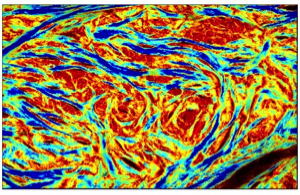
We have developed techniques to visualize the density and orientation of collagen fibers of soft tissues in high resolution (~um). Using these techniques we have measured microstructural properties of the tissues, such as the patterns of collagen crimp, that underlie the complex nonlinear properties of the eye. |
Wide field analysis of the eye architecture data has allowed us to propose new models of eye architecture. These models are then evaluated using advanced computational mechanics. |
We have developed a new generation of numerical models to study biomechanics of the eye. These models incorporate the unique information of eye architecture from our experiments. We are using them to identify the key factors that determine neural tissue sensitivity to mechanical insult and susceptibility to disease. |
| Whole-eye MRI | Microvasculature of the optic nerve head | Harnessing mechanostimulation to engineer corneal stroma |
 |
 |
 |
In collaboration with Kevin Chan (NYU) we are developing techniques to utilize magnetic resonance imaging to simultaneously characterize the structure, microarchitecture, mechanics and composition of the eye. These techniques will enable non-invasive quantification of the effects of aging and ocular hypertension. |
We work on a cross-species characterization of the vasculature of the posterior pole. Collaboration with Tatjana Jakobs and Joe Rizzo, Massachusetts Eye and Ear. |
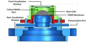
In collaboration with James Funderburgh (Pitt), we are working to engineer a corneal stroma graft suitable for transplant. |
| Novel tools for mechanical analysis of eye tissues | Non-invasive measurement of trabecular meshwork properties in vivo | |
 |
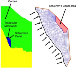 |
|
In collaboration with Zhongpin Chen (University of California at Irvine) and Qifa Zhou (University of Southern California) we are developing a novel elastographic system for use in the eye. The system integrates acoustic radiation force, phase sensitive optical coherence elastography and the analysis software to enable ex vivo and in vivo measurement of lamina cribrosa mechanical properties. |
In collaboration with Rouzbeh Amini (Akron) and Larry Kagemann (NYU and FDA), we are working to develop integrated imaging and analysis techniques to measure the mechanical properties of the trabecular meshwork in vivo. |
Financial Support
National Institutes of Health and National Eye Institute
Canadian Institutes of Health Research
Eye and Ear Foundation of Pittsburgh, PA
University of Pittsburgh SPRIG program
University of Pittsburgh CTSI program
Glaucoma Research Foundation