
Laboratory of
Ocular Biomechanics
University of Pittsburgh
Latest News
May/2025: Best wishes to Susannah!
Susannah was a PhD student then a post-doc in our lab. She has moved to Canada to be a post-doc at The University of Montreal. [Susannah's Publications]
May/2025: New paper accepted!
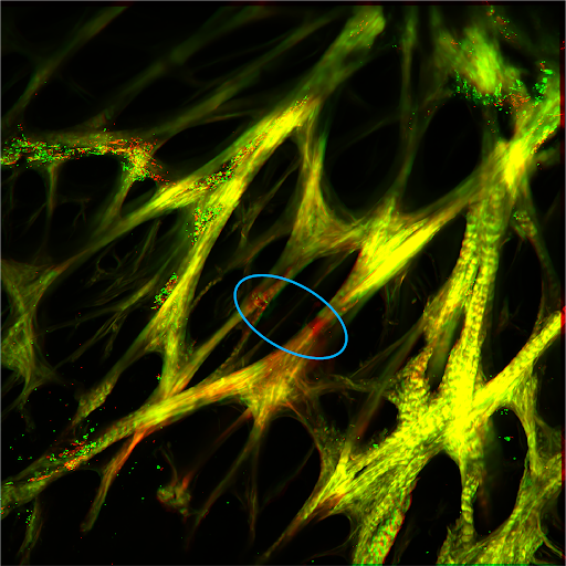
"Pericytes in the Optic Nerve Head" by Progress in Retinal and Eye Research. This work was led by Susannah Waxman from our lab, with a broad group of collaborators. [Read Here]
May/2025: Three talks and a poster!
We presented three talks (by Ian Sigal, Susannah Waxman, and Yuankai Lu) and a poster (by Bingrui Wang) at ARVO 2025 in Salt Lake City, Utah.
May/2025: Talk at ARVO Imaging!
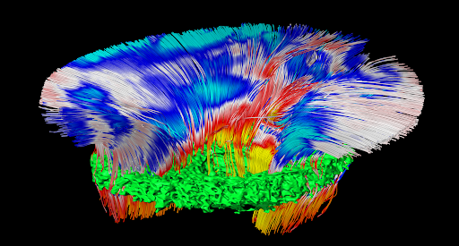
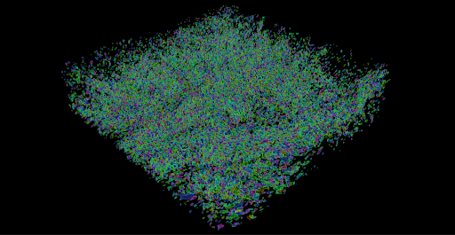
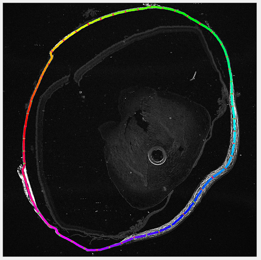
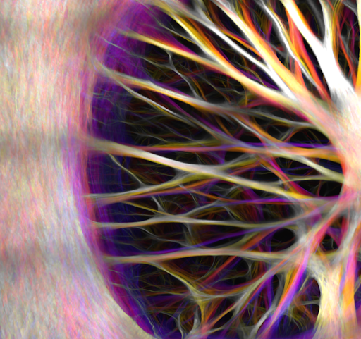
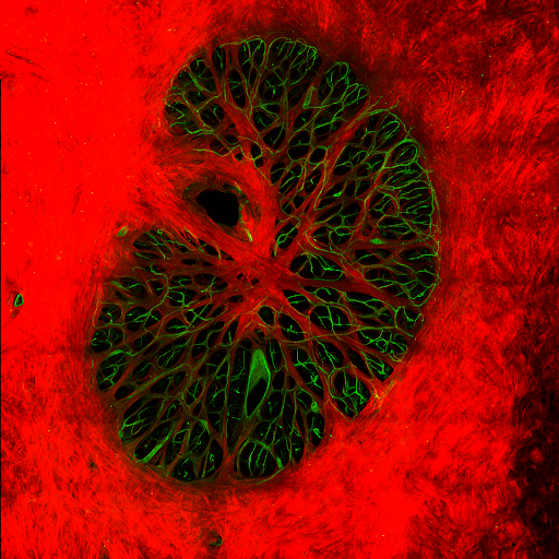
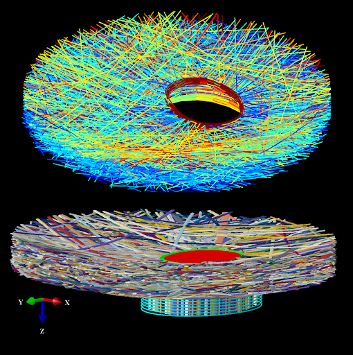
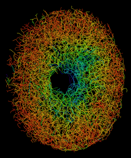
Susannah Waxman presented a talk titled, "High-resolution 3D imaging of the optically cleared ONH revals the intact structure of collagen and vascular networks" at ARVO Imaging 2025 in Salt Lake City, Utah.

Examples of our work


Why biomechanics of the eye?
|
In our daily lives we rarely think of the eye as a biomechanical structure. The eye, however, is a remarkably complex structure with biomechanics involved in many of its functions. For our eyes to be able to track moving objects, for example, requires a delicate balance of the forces exerted by several muscles. Forces are also responsible for deforming the lens and allow focusing. A slight imbalance between the forces and tissue properties may be enough to alter or even preclude vision. These effects may take place quickly or over long periods, even years. Understanding ocular biomechanics is therefore important for preventing and treating vision loss. |
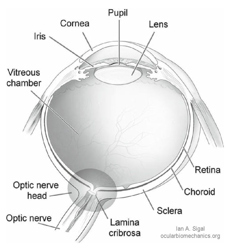
Schematic cross-section through a human eye. Light enters the eye through the cornea, passes through the pupil, lens and vitreous humour and strikes the retina, where it is absorbed. Retinal nerve fibers transmit visual information to the brain. These fibers converge at the optic nerve head region, exit the eye through the scleral canal, and form the optic nerve. The lamina cribrosa is a porous structure spanning the scleral canal. The vitreous chamber is filled with the vitreous humor, which exerts a pressure, the intraocular pressure, on the surface of the retina. [Sigal et al. Biomech Model Mechanobiol, 8(2):85-98, Apr 2009] (adapted from an illustration from NIH) |
Goals
The objective of the Laboratory of Ocular Biomechanics is to study the eye as a biomechanical structure. More specifically our work is aimed at identifying the causes of glaucoma, with the ultimate intention of finding a way to prevent vision loss.