
Laboratory of
Ocular Biomechanics
University of Pittsburgh
In-vivo/Ex-vivo Ocular biomechanics and microstructure
| Direct measurement of 3D deformations of the eye | Effects of aging | Interplay between intraocular and intracranial pressures |
 |
 |
 |
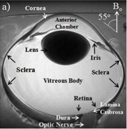
We have developed experimental and computational techniques to measure the acute and chronic displacements and deformations of the eye. |
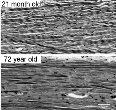
Age is one of the main risk factors for many diseases. We work to understand how aging affects the eye at the microstructural level. |
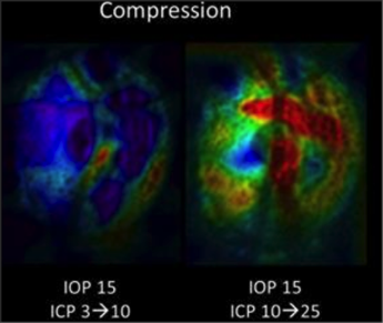
We have developed computational and experimental techniques to study the independent and interacting effects of intraocular and intracranial pressures on the optic nerve head. In collaboration with Gadi Wollstein (NYU) and Matthew Smith (Pitt). |
| Role of eye biomechanics on drusen | Novel imaging tools to study soft tissue collagen | Novel techniques for glaucoma diagnosis |
 |
 |
 |
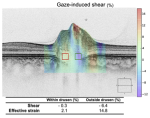
In collaboration with Patrick Sibony (Stonybrook), we are exploring the potential role of biomechanics on the progression of drusen. |
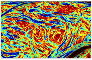
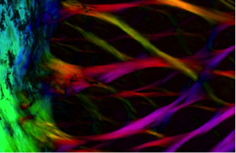
We have developed tools that enable measuring the full 3D orientation of collagen fibers. |
In collaboration with the Glaucoma imaging group (NYU), we are working to develop novel techniques for the diagnosis of glaucoma. |
| Microarchitecture of the eye | New models of eye architecture | Numerical models of eye biomechanics |
 |
 |
 |
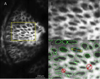
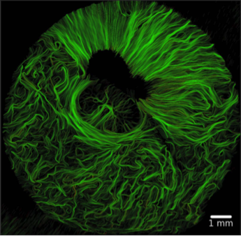
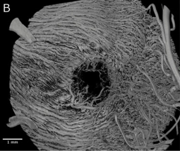
We have developed techniques to visualize the density and orientation of collagen fibers of soft tissues in high resolution (~um). Using these techniques we have measured microstructural properties of the tissues, such as the patterns of collagen crimp, that underlie the complex nonlinear properties of the eye. |
Wide field analysis of the eye architecture data has allowed us to propose new models of eye architecture. These models are then evaluated using advanced computational mechanics. |
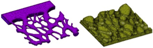
We have developed a new generation of numerical models to study biomechanics of the eye. These models incorporate the unique information of eye architecture from our experiments. We are using them to identify the key factors that determine neural tissue sensitivity to mechanical insult and susceptibility to disease. |
| Whole-eye MRI | Microvasculature of the optic nerve head | Harnessing mechanostimulation to engineer corneal stroma |
 |
 |
 |
In collaboration with Kevin Chan (NYU) we are developing techniques to utilize magnetic resonance imaging to simultaneously characterize the structure, microarchitecture, mechanics and composition of the eye. These techniques will enable non-invasive quantification of the effects of aging and ocular hypertension. |
We work on a cross-species characterization of the vasculature of the posterior pole. Collaboration with Tatjana Jakobs and Joe Rizzo, Massachusetts Eye and Ear. |
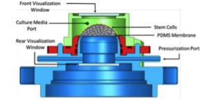
In collaboration with James Funderburgh (Pitt), we are working to engineer a corneal stroma graft suitable for transplant. |
| Novel tools for mechanical analysis of eye tissues | Non-invasive measurement of trabecular meshwork properties in vivo | |
 |
 |
|
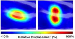
In collaboration with Zhongpin Chen (University of California at Irvine) and Qifa Zhou (University of Southern California) we are developing a novel elastographic system for use in the eye. The system integrates acoustic radiation force, phase sensitive optical coherence elastography and the analysis software to enable ex vivo and in vivo measurement of lamina cribrosa mechanical properties. |
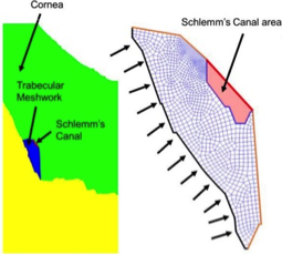
In collaboration with Rouzbeh Amini (Akron) and Larry Kagemann (NYU and FDA), we are working to develop integrated imaging and analysis techniques to measure the mechanical properties of the trabecular meshwork in vivo. |
Financial Support
National Institutes of Health and National Eye Institute
Canadian Institutes of Health Research
Eye and Ear Foundation of Pittsburgh, PA
University of Pittsburgh SPRIG program
University of Pittsburgh CTSI program
Glaucoma Research Foundation