Laboratory of
Ocular Biomechanics
University of Pittsburgh
Copyright Notice: The publishers hold the copyright of these articles. The PDFs are provided here to ensure rapid dissemination of scholarly work. It is understood that you will use them only in a manner consistent with the fair-use provisions of the relevant copyright laws. You may not distribute them or use them for any commercial enterprise.
bioRxiv Notice: Papers in biorXiv have not completed peer-review yet.
Publications
See publications of Dr. Sigal in PubMed, Google Scholar and Research Gate.


loading publications

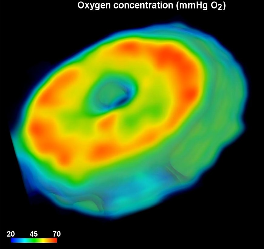
- Lamina cribrosa hypoxia sensitivity to variations of anatomy and vascular factors
- Accepted to ASME Journal of Biomechanical Engineering April 2025.
- [PDF]
- Lamina cribrosa hypoxia sensitivity to variations of anatomy and vascular factors
- Accepted to ASME Journal of Biomechanical Engineering April 2025.
- [PDF]
- [Supplementary Video]

- Impact of elevated IOP on lamina cribrosa oxygenation; A combined experimental-computational study on monkeys
- Ophthalmol Sci. 2025 Jan 31;5(3):100725. doi: 10.1016/j.xops.2025.100725. PMID: 40161464; PMCID: PMC11950774.
- [PDF,Link]
- [Supplementary Video 1,Supplementary Video 2, Supplementary Video 3]
- [Supplementary Video 4,Supplementary Video 5]


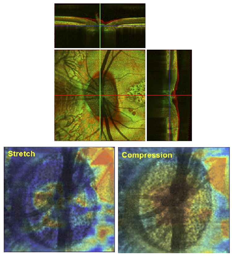
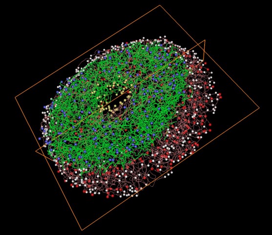
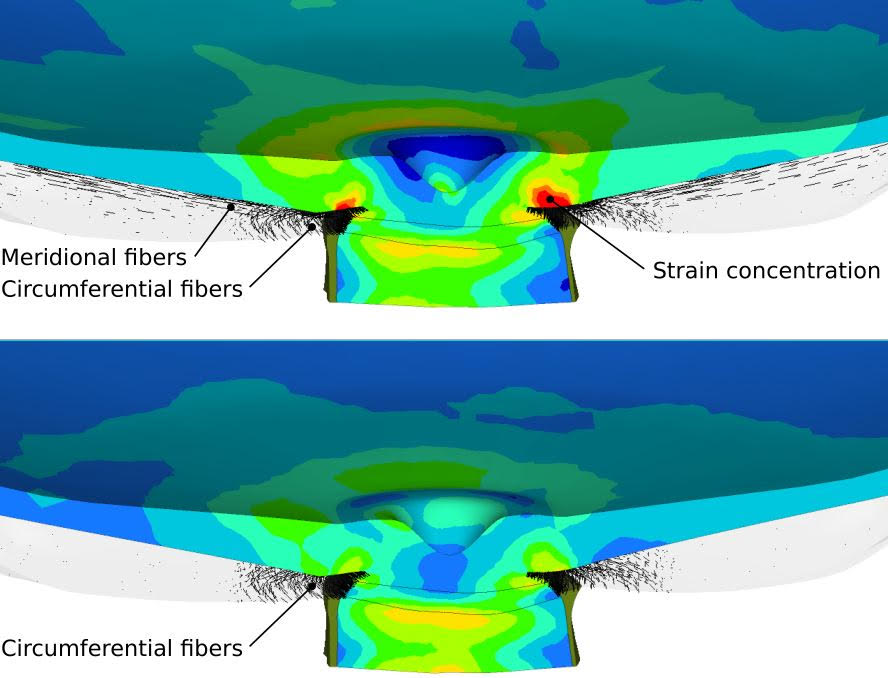
- Comparing continuum and direct fiber models of soft tissues. An ocular biomechanics example reveals that continuum models may artificially disrupt the strains at both the tissue and fiber levels
- Acta Biomater. 2024 Dec;190:317-328. doi: 10.1016/j.actbio.2024.10.019. Epub 2024 Oct 16. PMID: 39424020.
- [PDF,Link]


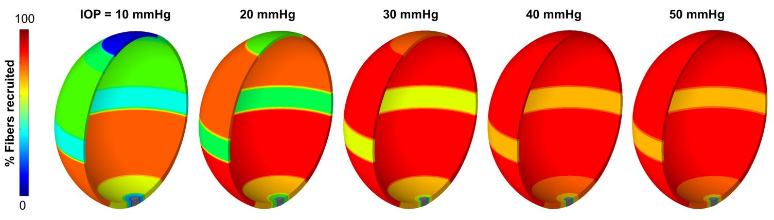
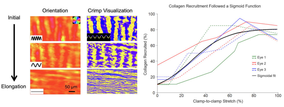
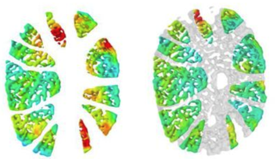
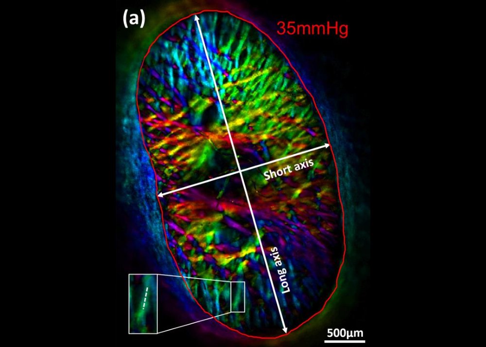
- Direct measurements of collagen fiber recruitment in the posterior pole of the eye
- Acta Biomaterialia 2024 Jan 1:173:135-147. doi: 10.1016/j.actbio.2023.11.013
- [PDF, Journal Link]
- Direct measurements of collagen fiber recruitment in the posterior pole of the eye
- Acta Biomaterialia 2024 Jan 1:173:135-147. doi: 10.1016/j.actbio.2023.11.013
- [PDF, Journal Link]
- 2D or not 2D? Mapping the in-depth inclination of the collagen fibers of the corneoscleral shell
- Experimental Eye Research 2023 Dec:237:109701. doi: 10.1016/j.exer.2023.109701
- [Link]
- 2D or not 2D? Mapping the in-depth inclination of the collagen fibers of the corneoscleral shell
- Experimental Eye Research 2023 Dec:237:109701. doi: 10.1016/j.exer.2023.109701
- [Link]
- So-called lamina cribrosa defects may mitigate IOP-induced neural tissue insult
- Investigative Ophthalmology and Visual Science, 61(15), November 2020.
- (* Authors contributed equally to this manuscript)
-
[PDF,
Free to read Link]
- So-called lamina cribrosa defects may mitigate IOP-induced neural tissue insult
- Investigative Ophthalmology and Visual Science, 61(15), November 2020.
- (* Authors contributed equally to this manuscript)
-
[PDF,
Free to read Link]
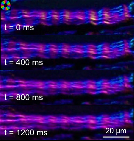
- Lamina cribrosa capillaries straighten as intraocular pressure increases
- Investigative Ophthalmology and Visual Science, 61(2), October 2020.
-
[PDF,
Free to read Link]
- Lamina cribrosa capillaries straighten as intraocular pressure increases
- Investigative Ophthalmology and Visual Science, 61(2), October 2020.
-
[PDF,
Free to read Link]


- Radial and circumferential collagen fibers are a feature of the peripapillary sclera of human, monkey, pig, cow, goat and sheep
- Investigative Ophthalmology and Visual Science, 59(12), 4763-4774, October 2018.
-
[PDF,
Free to read Link]
- Radial and circumferential collagen fibers are a feature of the peripapillary sclera of human, monkey, pig, cow, goat and sheep
- Investigative Ophthalmology and Visual Science, 59(12), 4763-4774, October 2018.
-
[PDF,
Free to read Link]
- Thin lamina cribrosa beams have different collagen microstructure than thick beams
- Investigative Ophthalmology and Visual Science, 59(11), 4653-4661, September 2018.
- (* Authors contributed equally to this manuscript)
-
[PDF,
Free to read Link]
- Thin lamina cribrosa beams have different collagen microstructure than thick beams
- Investigative Ophthalmology and Visual Science, 59(11), 4653-4661, September 2018.
- (* Authors contributed equally to this manuscript)
-
[PDF,
Free to read Link]

- Spatial patterns and age-related changes of the collagen crimp in the human cornea and sclera
- Investigative Ophthalmology and Visual Science, 59(7), 2987-2998, June 2018.
-
[PDF,
Free to read Link]
- Spatial patterns and age-related changes of the collagen crimp in the human cornea and sclera
- Investigative Ophthalmology and Visual Science, 59(7), 2987-2998, June 2018.
-
[PDF,
Free to read Link]
- Tortuous pore path through the glaucomatous lamina cribrosa
- Scientific reports, 8(1), 7281, May 2018.
-
[PDF,
Free to read link]
- Tortuous pore path through the glaucomatous lamina cribrosa
- Scientific reports, 8(1), 7281, May 2018.
-
[PDF,
Free to read link]

- Seeing the hidden lamina; Effects of exsanguination on the optic nerve head
- Investigative Ophthalmology and Visual Science, 59, 2564-2575, May 2018.
-
[PDF,
Free to read Link]
- Seeing the hidden lamina; Effects of exsanguination on the optic nerve head
- Investigative Ophthalmology and Visual Science, 59, 2564-2575, May 2018.
-
[PDF,
Free to read Link]
- Cerebrospinal Fluid Pressure; Revisiting Factors Influencing Optic Nerve Head Biomechanics
- Investigative Ophthalmology and Visual Science, 59(1), 154-165, January 2018.
-
[PDF,
Free to read Link]
- Cerebrospinal Fluid Pressure; Revisiting Factors Influencing Optic Nerve Head Biomechanics
- Investigative Ophthalmology and Visual Science, 59(1), 154-165, January 2018.
-
[PDF,
Free to read Link]
- Lamina Cribrosa Pore Shape and Size as Predictors of Neural Tissue Mechanical Insult
- Investigative Ophthalmology and Visual Science, 58(12), 5336-5346, October 2017.
-
[PDF,
Free to read Link]
- Lamina Cribrosa Pore Shape and Size as Predictors of Neural Tissue Mechanical Insult
- Investigative Ophthalmology and Visual Science, 58(12), 5336-5346, October 2017.
-
[PDF,
Free to read Link]
- Formalin Fixation and Cryosectioning Cause Only Minimal Changes in Shape or Size of Ocular Tissues
- Scientific Reports, 7(1), 12065, September 2017.
-
[PDF,
Free to read link]
- Formalin Fixation and Cryosectioning Cause Only Minimal Changes in Shape or Size of Ocular Tissues
- Scientific Reports, 7(1), 12065, September 2017.
-
[PDF,
Free to read link]

- Microstructural Crimp of the Lamina Cribrosa and Peripapillary Sclera Collagen Fibers
- Investigative Ophthalmology and Visual Science, 58(9), 3378-3388, July 2017.
-
[PDF,
Free to read link]
- Microstructural Crimp of the Lamina Cribrosa and Peripapillary Sclera Collagen Fibers
- Investigative Ophthalmology and Visual Science, 58(9), 3378-3388, July 2017.
-
[PDF,
Free to read link]
- Thick Prelaminar Tissue Decreases Lamina Cribrosa Visibility
- Investigative Ophthalmology and Visual Science, 58(3), 1751-1757, March 2017.
-
[PDF,
Free to read link]
- Thick Prelaminar Tissue Decreases Lamina Cribrosa Visibility
- Investigative Ophthalmology and Visual Science, 58(3), 1751-1757, March 2017.
-
[PDF,
Free to read link]
- An imaged-based inverse finite element method to determine in-vivo mechanical properties of human trabecular meshwork
- Journal for Modeling in Ophthalmology, 1(3), 100-111, 2017.
-
[PDF,
Free to read link]
- An imaged-based inverse finite element method to determine in-vivo mechanical properties of human trabecular meshwork
- Journal for Modeling in Ophthalmology, 1(3), 100-111, 2017.
-
[PDF,
Free to read link]

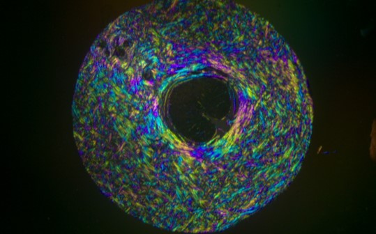
- Collagen architecture of the posterior pole; high-resolution, wide-field-of-view visualization and analysis using polarized light microscopy
- Investigative Ophthalmology and Visual Science, 58(2), 735744, February 2017.
-
[PDF,
Free to read link]
- Collagen architecture of the posterior pole; high-resolution, wide-field-of-view visualization and analysis using polarized light microscopy
- Investigative Ophthalmology and Visual Science, 58(2), 735744, February 2017.
-
[PDF,
Free to read link]
- Non-invasive MRI Assessments of Tissue Microstructures and Macromolecules in the Eye upon Biomechanical or Biochemical Modulation
- Scientific Reports, 6(1), 1-14, August 2016.
-
[PDF,
Free to read link]
- Non-invasive MRI Assessments of Tissue Microstructures and Macromolecules in the Eye upon Biomechanical or Biochemical Modulation
- Scientific Reports, 6(1), 1-14, August 2016.
-
[PDF,
Free to read link]
- Experimental glaucoma causes optic nerve head neural rim tissue compression: a potentially important mechanism of axon injury
- Investigative Ophthalmology and Visual Science, 57(10), 4403-4411, August 2016.
-
[PDF,
Free to read link]
- Experimental glaucoma causes optic nerve head neural rim tissue compression: a potentially important mechanism of axon injury
- Investigative Ophthalmology and Visual Science, 57(10), 4403-4411, August 2016.
-
[PDF,
Free to read link]
- Decreased lamina cribrosa beam thickness and pore diameter relative to distance from the central retinal vessel trunk
- Investigative Ophthalmology and Visual Science, 57(7), 3088-3092, June 2016.
-
[PDF,
Free to read link]
- Decreased lamina cribrosa beam thickness and pore diameter relative to distance from the central retinal vessel trunk
- Investigative Ophthalmology and Visual Science, 57(7), 3088-3092, June 2016.
-
[PDF,
Free to read link]
- Regionally Discrete Aqueous Humor Outflow Quantification Using Fluorescein Canalograms
- PLoS ONE, 11(3), e0151754, March 2016.
-
[PDF,
Free to read link]
- Regionally Discrete Aqueous Humor Outflow Quantification Using Fluorescein Canalograms
- PLoS ONE, 11(3), e0151754, March 2016.
-
[PDF,
Free to read link]
- A Problem of Proportions in OCT-based Morphometry and a Proposed Solution
- Investigative Ophthalmology and Visual Science, 57(2), 484-485, Feburary 2016.
- (Letter to the Editor)
-
[PDF,
Free to read link]
- A Problem of Proportions in OCT-based Morphometry and a Proposed Solution
- Investigative Ophthalmology and Visual Science, 57(2), 484-485, Feburary 2016.
- (Letter to the Editor)
-
[PDF,
Free to read link]

- Use and Misuse of Laplace’s Law in Ophthalmology
- Investigative Ophthalmology and Visual Science, 57(1), 236-245, January 2016.
-
[PDF,
Free to read link]
- Use and Misuse of Laplace’s Law in Ophthalmology
- Investigative Ophthalmology and Visual Science, 57(1), 236-245, January 2016.
-
[PDF,
Free to read link]

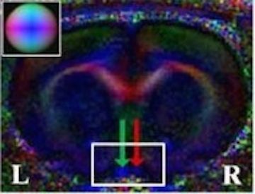
- In Vivo Evaluation of White Matter Inegrity and Anterograde Transport in Visual Systems After Excitoxic Retinal Injury with Multimodal MRI and OCT
- Investigative Ophthalmology and Visual Science, 56(6), 3788-3800, June 2015.
-
[PDF,
Free to read link]
- In Vivo Evaluation of White Matter Inegrity and Anterograde Transport in Visual Systems After Excitoxic Retinal Injury with Multimodal MRI and OCT
- Investigative Ophthalmology and Visual Science, 56(6), 3788-3800, June 2015.
-
[PDF,
Free to read link]
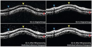
- Histogram Matching Extends Acceptable Signal Strength Range on Optical Coherence Tomography Images
- Investigative Ophthalmology and Visual Science, 56(6), 3810-3819, June 2015.
-
[PDF,
Free to read link]
- Histogram Matching Extends Acceptable Signal Strength Range on Optical Coherence Tomography Images
- Investigative Ophthalmology and Visual Science, 56(6), 3810-3819, June 2015.
-
[PDF,
Free to read link]
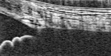
- Trabecular Meshwork Response to Pressure Elevation in the Living Human Eye
- Journal of Visualized Experiments, 20(100), e52611, June 2015.
- [Link]
- Trabecular Meshwork Response to Pressure Elevation in the Living Human Eye
- Journal of Visualized Experiments, 20(100), e52611, June 2015.
- [Link]
- In Vivo Three-Dimensional Characterization of the Healthy Human Lamina Cribrosa with Adaptive Optics Spectral-Domain Optical Coherence Tomography
- Investigative Ophthalmology and Visual Science, 55(10), 6459-6466, October 2014.
-
[PDF,
Free to read link]
- In Vivo Three-Dimensional Characterization of the Healthy Human Lamina Cribrosa with Adaptive Optics Spectral-Domain Optical Coherence Tomography
- Investigative Ophthalmology and Visual Science, 55(10), 6459-6466, October 2014.
-
[PDF,
Free to read link]
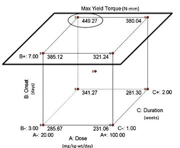
- Magic Angle-Enhanced MRI Of Fibrous Microstructures In Sclera And Cornea With And Without Intraocular Pressure Loading
- Investigative Ophthalmology and Visual Science, 55(9), 5662-5672, September 2014.
- (* Authors contributed equally to this manuscript)
-
[PDF,
Free to read link]
- Magic Angle-Enhanced MRI Of Fibrous Microstructures In Sclera And Cornea With And Without Intraocular Pressure Loading
- Investigative Ophthalmology and Visual Science, 55(9), 5662-5672, September 2014.
- (* Authors contributed equally to this manuscript)
-
[PDF,
Free to read link]

- Recent Advances in OCT Imaging of the Lamina Cribrosa
- British Journal of Ophthalmology, 98(Suppl 2), ii34-ii39, July 2014.
-
[PDF,
Free to read link]
- Recent Advances in OCT Imaging of the Lamina Cribrosa
- British Journal of Ophthalmology, 98(Suppl 2), ii34-ii39, June 2014.
-
[PDF,
Free to read link]
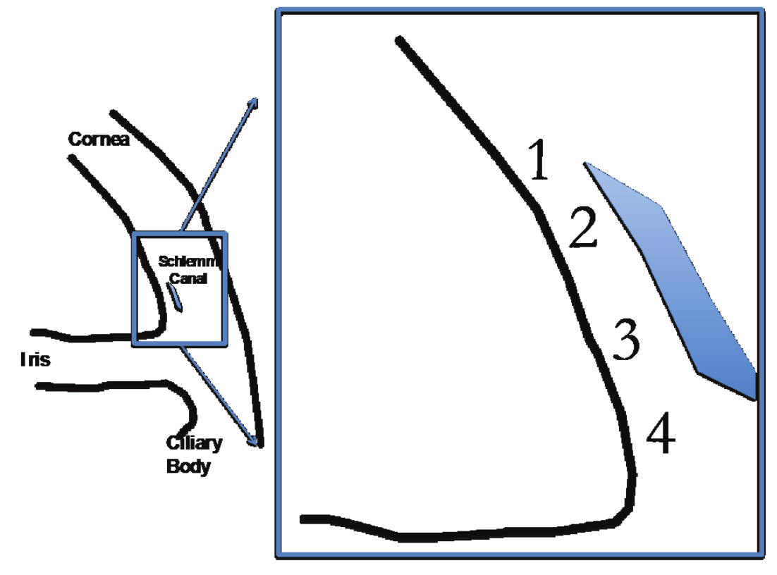
- Characterization of Schlemm’s Canal Cross-Sectional Area
- British Journal of Ophthalmology, 98(Suppl 2), ii10-ii14, June 2014.
-
[PDF,
Free to read link]
- Characterization of Schlemm’s Canal Cross-Sectional Area
- British Journal of Ophthalmology, 98(Suppl 2), ii10-ii14, June 2014.
-
[PDF,
Free to read link]
- IOP Elevation Reduces Schlemm’s Canal Cross-sectional Area
- Investigative Ophthalmology and Visual Science, 55(3), 1805-1809, March 2014.
-
[PDF,
Free to read link]
- IOP Elevation Reduces Schlemm’s Canal Cross-sectional Area
- Investigative Ophthalmology and Visual Science, 55(3), 1805-1809, March 2014.
-
[PDF,
Free to read link]
- Gold Nanorods as a Contrast Agent for Doppler Optical Coherence Tomography
- PLoS One, 9(3), e90690, March 2014.
-
[PDF,
Free to read link]
- Gold Nanorods as a Contrast Agent for Doppler Optical Coherence Tomography
- PLoS One, 9(3), e90690, March 2014.
-
[PDF,
Free to read link]

- Eye-Specific IOP-Induced Displacements and Deformations of Human Lamina Cribrosa
- Investigative Ophthalmology and Visual Science, 55(1), 1-15, January 2014.
-
[PDF,
Free to read link]
- eCover of the January 2014 issue of IOVS
- Research highlight by Thao D. Nguyen of Johns Hopkins:
- "A Significant Advance in the Biomechanical Evaluation of the Optic Nerve Head"
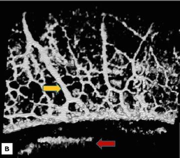
- In-Vivo Lamina Cribrosa Microarchitecture in Healthy and Glaucomatous Eyes as Assessed by Optical Coherence Tomography
- Investigative Ophthalmology and Visual Science, 54(13), 8270-8274, December 2013.
-
[PDF,
Free to read link]
- In-Vivo Lamina Cribrosa Microarchitecture in Healthy and Glaucomatous Eyes as Assessed by Optical Coherence Tomography
- Investigative Ophthalmology and Visual Science, 54(13), 8270-8274, December 2013.
-
[PDF,
Free to read link]
- Signal Normalization Reduces Systematic Measurement Differences Between Spectral Domain Optical Coherence Tomography Devices
- Investigative Ophthalmology and Visual Science, 54(12), 7317-7322, November 2013.
-
[PDF,
Free to read link]
- Signal Normalization Reduces Systematic Measurement Differences Between Spectral Domain Optical Coherence Tomography Devices
- Investigative Ophthalmology and Visual Science, 54(12), 7317-7322, November 2013.
-
[PDF,
Free to read link]
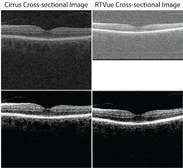
- Individual A-Scan Signal Normalization Between Two Spectral Domain Optical Coherence Tomography Devices
- Investigative Ophthalmology and Visual Science, 54(5),3463-3471, May 2013.
-
[PDF,
Free to read link]
- Individual A-Scan Signal Normalization Between Two Spectral Domain Optical Coherence Tomography Devices
- Investigative Ophthalmology and Visual Science, 54(5), 3463-3471, May 2013.
-
[PDF,
Free to read link]
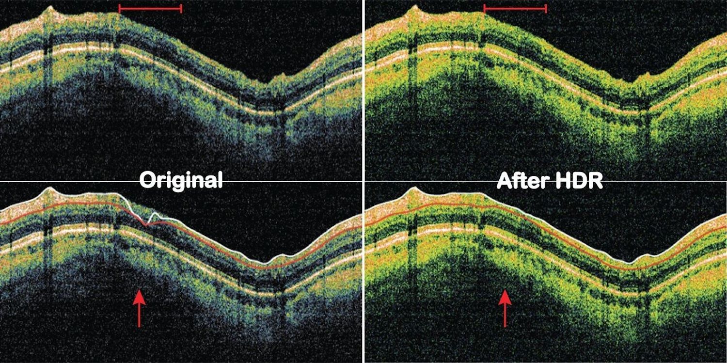
- High Dynamic Range Imaging Concept-Based Signal Enhancement Method Reduced the Optical Coherence Tomography Measurement Variability
- Investigative Ophthalmology and Visual Science, 54(1), 836-841, January 2013.
-
[PDF,
Free to read link]
- High Dynamic Range Imaging Concept-Based Signal Enhancement Method Reduced the Optical Coherence Tomography Measurement Variability
- Investigative Ophthalmology and Visual Science, 54(1):836-41, Jan 2013. PMID 23299477.
-
[PDF,
Free to read link]
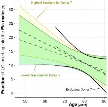
- Human Lamina Cribrosa Insertion and Age
- Investigative Ophthalmology and Visual Science, 53(11), 6780-6789, October 2012.
-
[PDF,
Free to read link]
- Human Lamina Cribrosa Insertion and Age
- Investigative Ophthalmology and Visual Science, 53(11):6780-9, Oct 2012. PMID 22956611.
-
[PDF,
Free to read link]
- Morphometric Analysis of Aqueous Humor Outflow Structures with Spectral Domain Optical Coherence Tomography
- Investigative Ophthalmology and Visual Science, 53(9), 5198-207, September 2012.
-
[PDF,
Free to read link]
- Morphometric Analysis of Aqueous Humor Outflow Structures with Spectral Domain Optical Coherence Tomography
- Invest Opthalmol Vis Sci, 53(9):5198-207, September 2012.
-
[PDF,
Free to read link]

- A few good responses. Which mechanical effects of IOP on the ONH to study?
- Investigative Ophthalmology and Visual Science, 53(7), 4270-4278, June 2012.
-
[PDF,
Free to read link]
- A few good responses. Which mechanical effects of IOP on the ONH to study?
- Investigative Ophthalmology and Visual Science, 53(7), 4270-4278, June 2012.
-
[PDF,
Free to read link]
- The Optic Nerve Head As A Robust Biomechanical System
- Investigative Ophthalmology and Visual Science, 53(6), 2658-2667, May 2012.
-
[PDF,
Free to read link]
- The Optic Nerve Head As A Robust Biomechanical System
- Investigative Ophthalmology and Visual Science, 53(6), 2658-2667, May 2012.
-
[PDF,
Free to read link]
- Effect of Acute Intraocular Pressure Elevation on the Monkey Optic Nerve Head as Detected by Spectral Domain Ocular Coherence Tomography
- Investigative Ophthalmology and Visual Science, 52(12), 9431-9437, December 2011.
-
[PDF,
Free to read link]
- Effect of Acute Intraocular Pressure Elevation on the Monkey Optic Nerve Head as Detected by Spectral Domain Ocular Coherence Tomography
- Investigative Ophthalmology and Visual Science, 52(12), 9431-9437, December 2011.
-
[PDF,
Free to read link]

- IOP-induced lamina cribrosa displacement and scleral canal expansion. Are they independent or related?
- Investigative Ophthalmology and Visual Science, 52(12), 9023-9032. December 2011.
-
[PDF,
Free to read link]
- IOP-induced lamina cribrosa displacement and scleral canal expansion. Are they independent or related?
- Investigative Ophthalmology and Visual Science, 52(12), 9023-9032. December 2011.
-
[PDF,
Free to read link]
- Posterior (outward) migration of the lamina cribrosa and early cupping in monkey experimental glaucoma
- Investigative Ophthalmology and Visual Science, 52(10), 7109-7921, September 2011.
-
[PDF,
Free to read link]
- Posterior (outward) migration of the lamina cribrosa and early cupping in monkey experimental glaucoma
- Investigative Ophthalmology and Visual Science, 52(10), 7109-7921, September 2011.
-
[PDF,
Free to read link]
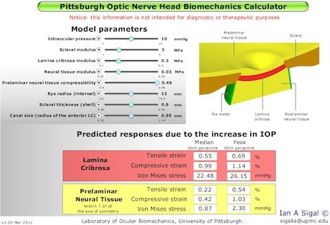
- An Applet to Estimate the IOP-Induced Stress and Strain Within the Optic Nerve Head
- Investigative Ophthalmology and Visual Science, 52(8), 5497-5506, July 2011.
-
[PDF,
Free to read link]
- Note: The applet associated with this paper is in the Software page.
- An Applet to Estimate the IOP-Induced Stress and Strain Within the Optic Nerve Head
- Investigative Ophthalmology and Visual Science, 52(8), 5497-5506, July 2011.
-
[PDF,
Free to read link]
- Note: The applet associated with this paper is in the Software page.
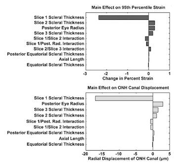
- IOP-Induced Lamina Cribrosa Displacement and Scleral Canal Expansion: an Analysis of Factor Interactions Using Parameterized Eye-Specific Models
- Investigative Ophthalmology and Visual Science, 52(3), 1896-1907, March 2011.
-
[PDF,
Free to read link]
- IOP-Induced Lamina Cribrosa Displacement and Scleral Canal Expansion: an Analysis of Factor Interactions Using Parameterized Eye-Specific Models
- Investigative Ophthalmology and Visual Science, 52(3), 1896-1907, March 2011.
-
[PDF,
Free to read link]
- Longitudinal Change Detected by Spectral Domain Optical Coherence Tomography in the Glaucomatous Optic Nerve Head and Peripapillary Retina
- Investigative Ophthalmology and Visual Science, 52(3), 1206-1219, March 2011.
-
[PDF,
Free to read link]
- Longitudinal Change Detected by Spectral Domain Optical Coherence Tomography in the Glaucomatous Optic Nerve Head and Peripapillary Retina
- Investigative Ophthalmology and Visual Science, 52(3), 1206-19, March 2011.
-
[PDF,
Free to read link]
- Deformation of the Early Glaucomatous Monkey Optic Nerve Head Connective Tissue Following Acute IOP Elevation Within 3-D Histomorphometric Reconstructions
- Investigative Ophthalmology and Visual Science, 52(1), 345-363, January 2011.
-
[PDF,
Free to read link]
- Deformation of the Early Glaucomatous Monkey Optic Nerve Head Connective Tissue Following Acute IOP Elevation Within 3-D Histomorphometric Reconstructions
- Investigative Ophthalmology and Visual Science, 52(1), 345-363, January 2011.
-
[PDF,
Free to read link]
- Changes in the Biomechanical Response of the Optic Nerve Head in Early Experimental Glaucoma
- Investigative Ophthalmology and Visual Science, 51(11):5675-5684. November 2010, PMID 20538997
-
[PDF,
Free to read link]
- Changes in the Biomechanical Response of the Optic Nerve Head in Early Experimental Glaucoma
- Investigative Ophthalmology and Visual Science, 51(11):5675-5684. November 2010, PMID 20538997
-
[PDF,
Free to read link]

- Biomechanical Changes of the Optic Disc
- Ocular Disease: Mechanisms and Management Eds: LA Levin and DM Albert, Saunders (Elsevier), Chapter 20, 704, March 2010.
- ISBN 978-0-7020-2983-7
- [PDF]
- Biomechanical Changes of the Optic Disc
- Ocular Disease: Mechanisms and Management Eds: LA Levin and DM Albert, Saunders (Elsevier), Chapter 20, 704, March 2010.
- ISBN 978-0-7020-2983-7
- [PDF]


- Correlation Between Local Stress and Strain and Lamina Cribrosa Connective Tissue Volume Fraction in Normal Monkey Eyes
- Investigative Ophthalmology and Visual Science, 51(1), 295-307, January 2010.
-
[PDF,
Free to read link]
- Correlation Between Local Stress and Strain and Lamina Cribrosa Connective Tissue Volume Fraction in Normal Monkey Eyes
- Investigative Ophthalmology and Visual Science, 51(1), 295-307, January 2010.
-
[PDF,
Free to read link]
- Deformation of the Normal Monkey Optic Nerve Head Connective Tissues Following Acute IOP Elevation Within 3-D Histomorphometric Reconstructions
- Investigative Ophthalmology and Visual Science, 50(12), 5785-5789. December 2009.
-
[PDF,
Free to read link]
- Deformation of the Normal Monkey Optic Nerve Head Connective Tissues Following Acute IOP Elevation Within 3-D Histomorphometric Reconstructions
- Investigative Ophthalmology and Visual Science, 50(12), 5785-5789. December 2009.
-
[PDF,
Free to read link]
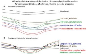
- Interactions Between Geometry and Mechanical Properties on the Optic Nerve Head
- Investigative Ophthalmology and Visual Science, 50(6), 2785-2795, June 2009.
-
[PDF,
Free to read link]
- Interactions Between Geometry and Mechanical Properties on the Optic Nerve Head
- Investigative Ophthalmology and Visual Science, 50(6), 2785-2795, June 2009.
-
[PDF,
Free to read link]
- Factors Influencing Human Optic Nerve Head Biomechanics
- Investigative Ophthalmology and Visual Science, 46(11), 4189-4199, November 2005.
-
[PDF,
Free to read link]
- Factors Influencing Human Optic Nerve Head Biomechanics
- Investigative Ophthalmology and Visual Science, 46(11), 4189-4199, November 2005.
-
[PDF,
Free to read link]

- Finite Element Modeling of Optic Nerve Head Biomechanics
- Investigative Ophthalmology and Visual Science, 45(12), 4378-4387, December 2004.
-
[PDF,
Free to read link]
- Finite Element Modeling of Optic Nerve Head Biomechanics
- Investigative Ophthalmology and Visual Science, 45(12), 4378-4387, December 2004.
-
[PDF,
Free to read link]

Publications
See publications of Dr. Sigal in PubMed, Google Scholar and Research Gate.
loading publications
- Lamina cribrosa hypoxia sensitivity to variations of anatomy and vascular factors
- Accepted to ASME Journal of Biomechanical Engineering April 2025.
- [PDF]
|
|
|
|
|
|
|
|
- Comparing continuum and direct fiber models of soft tissues. An ocular biomechanics example reveals that continuum models may artificially disrupt the strains at both the tissue and fiber levels
- Acta Biomater. 2024 Dec;190:317-328. doi: 10.1016/j.actbio.2024.10.019. Epub 2024 Oct 16. PMID: 39424020.
- [PDF,Link]
|
|
|
|
|
|
|
|
|
|
|
- Direct measurements of collagen fiber recruitment in the posterior pole of the eye
- Acta Biomaterialia 2024 Jan 1:173:135-147. doi: 10.1016/j.actbio.2023.11.013
- [PDF, Journal Link]
|
|
|
- 2D or not 2D? Mapping the in-depth inclination of the collagen fibers of the corneoscleral shell
- Experimental Eye Research 2023 Dec:237:109701. doi: 10.1016/j.exer.2023.109701
- [Link]
|
|
|
|
|
|
|
|
|
|
|
|
|
|
|
|
|
|
|
|
|
|
|
|
|
|
|
|
|
|
|
|
|
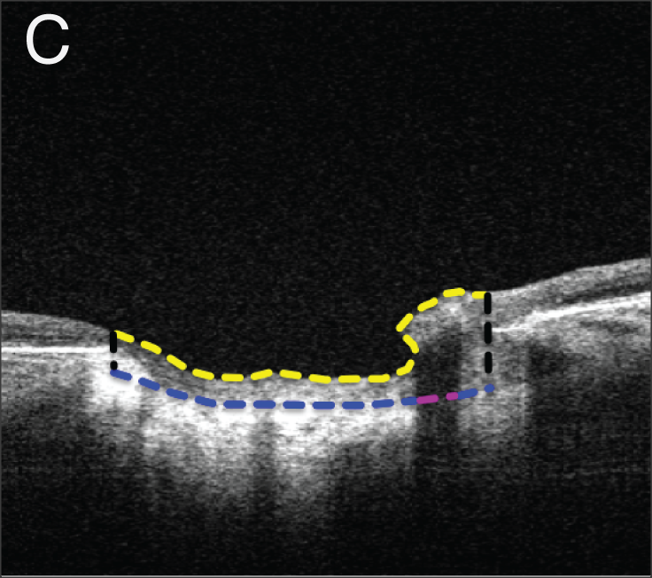
- So-called lamina cribrosa defects may mitigate IOP-induced neural tissue insult
- Investigative Ophthalmology and Visual Science, 61(15), November 2020.
- (* Authors contributed equally to this manuscript)
- [PDF, Free to read Link]
|
|
|
- Lamina cribrosa capillaries straighten as intraocular pressure increases
- Investigative Ophthalmology and Visual Science, 61(2), October 2020.
- [PDF, Free to read Link]
|
|
|
|
|
|
|
|
|
|
|
|
|
|
|
|
|
|
|
|
|
- Radial and circumferential collagen fibers are a feature of the peripapillary sclera of human, monkey, pig, cow, goat and sheep
- Investigative Ophthalmology and Visual Science, 59(12), 4763-4774, October 2018.
- [PDF, Free to read Link]
|
|
|
- Thin lamina cribrosa beams have different collagen microstructure than thick beams
- Investigative Ophthalmology and Visual Science, 59(11), 4653-4661, September 2018.
- (* Authors contributed equally to this manuscript)
- [PDF, Free to read Link]
|
|
|
|
|
- Spatial patterns and age-related changes of the collagen crimp in the human cornea and sclera
- Investigative Ophthalmology and Visual Science, 59(7), 2987-2998, June 2018.
- [PDF, Free to read Link]
|
|
|
- Tortuous pore path through the glaucomatous lamina cribrosa
- Scientific reports, 8(1), 7281, May 2018.
- [PDF, Free to read link]
|
|
|
|
|
|
|
|
|
|
|
- Seeing the hidden lamina; Effects of exsanguination on the optic nerve head
- Investigative Ophthalmology and Visual Science, 59, 2564-2575, May 2018.
- [PDF, Free to read Link]
|
|
|
|
|
|
|
- Cerebrospinal Fluid Pressure; Revisiting Factors Influencing Optic Nerve Head Biomechanics
- Investigative Ophthalmology and Visual Science, 59(1), 154-165, January 2018.
- [PDF, Free to read Link]
|
|
|
|
|
- Lamina Cribrosa Pore Shape and Size as Predictors of Neural Tissue Mechanical Insult
- Investigative Ophthalmology and Visual Science, 58(12), 5336-5346, October 2017.
- [PDF, Free to read Link]
|
|
|
- Formalin Fixation and Cryosectioning Cause Only Minimal Changes in Shape or Size of Ocular Tissues
- Scientific Reports, 7(1), 12065, September 2017.
- [PDF, Free to read link]
|
|
|
|
|
|
|
|
|
- Microstructural Crimp of the Lamina Cribrosa and Peripapillary Sclera Collagen Fibers
- Investigative Ophthalmology and Visual Science, 58(9), 3378-3388, July 2017.
- [PDF, Free to read link]
|
|
|
- Thick Prelaminar Tissue Decreases Lamina Cribrosa Visibility
- Investigative Ophthalmology and Visual Science, 58(3), 1751-1757, March 2017.
- [PDF, Free to read link]
|
|
|
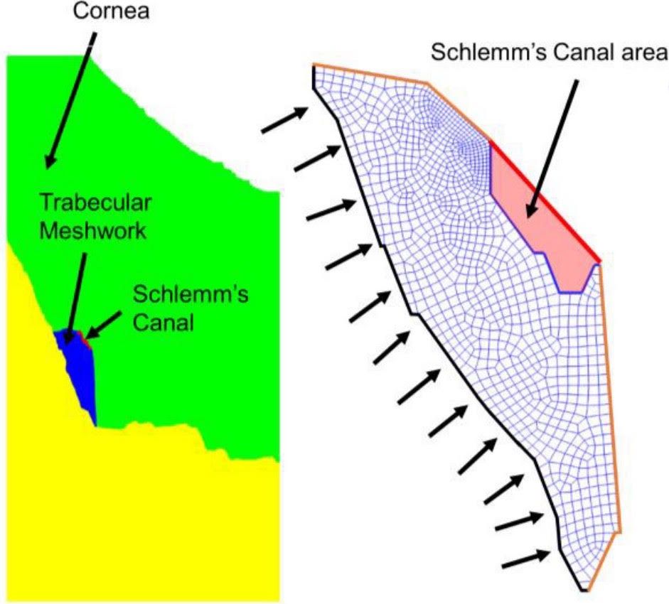
- An imaged-based inverse finite element method to determine in-vivo mechanical properties of human trabecular meshwork
- Journal for Modeling in Ophthalmology, 1(3), 100-111, 2017.
- [PDF, Free to read link]
|
|
|
|
|
|
|
|
|
|
|
- Collagen architecture of the posterior pole; high-resolution, wide-field-of-view visualization and analysis using polarized light microscopy
- Investigative Ophthalmology and Visual Science, 58(2), 735744, February 2017.
- [PDF, Free to read link]
|
|
|
|
|
- Non-invasive MRI Assessments of Tissue Microstructures and Macromolecules in the Eye upon Biomechanical or Biochemical Modulation
- Scientific Reports, 6(1), 1-14, August 2016.
- [PDF, Free to read link]
|
|
|
- Experimental glaucoma causes optic nerve head neural rim tissue compression: a potentially important mechanism of axon injury
- Investigative Ophthalmology and Visual Science, 57(10), 4403-4411, August 2016.
- [PDF, Free to read link]
|
|
|
|
|
- Decreased lamina cribrosa beam thickness and pore diameter relative to distance from the central retinal vessel trunk
- Investigative Ophthalmology and Visual Science, 57(7), 3088-3092, June 2016.
- [PDF, Free to read link]
|
|
|
- Regionally Discrete Aqueous Humor Outflow Quantification Using Fluorescein Canalograms
- PLoS ONE, 11(3), e0151754, March 2016.
- [PDF, Free to read link]
|
|
|
- A Problem of Proportions in OCT-based Morphometry and a Proposed Solution
- Investigative Ophthalmology and Visual Science, 57(2), 484-485, Feburary 2016.
- (Letter to the Editor)
- [PDF, Free to read link]
|
|
|
|
|
- Use and Misuse of Laplace’s Law in Ophthalmology
- Investigative Ophthalmology and Visual Science, 57(1), 236-245, January 2016.
- [PDF, Free to read link]
|
|
|
|
|
|
|
|
|
- In Vivo Evaluation of White Matter Inegrity and Anterograde Transport in Visual Systems After Excitoxic Retinal Injury with Multimodal MRI and OCT
- Investigative Ophthalmology and Visual Science, 56(6), 3788-3800, June 2015.
- [PDF, Free to read link]
|
|
|
- Histogram Matching Extends Acceptable Signal Strength Range on Optical Coherence Tomography Images
- Investigative Ophthalmology and Visual Science, 56(6), 3810-3819, June 2015.
- [PDF, Free to read link]
|
|
|
- Trabecular Meshwork Response to Pressure Elevation in the Living Human Eye
- Journal of Visualized Experiments, 20(100), e52611, June 2015.
- [Link]
|
|
|
|
|
|
|
- In Vivo Three-Dimensional Characterization of the Healthy Human Lamina Cribrosa with Adaptive Optics Spectral-Domain Optical Coherence Tomography
- Investigative Ophthalmology and Visual Science, 55(10), 6459-6466, October 2014.
- [PDF, Free to read link]
|
|
|
- Magic Angle-Enhanced MRI Of Fibrous Microstructures In Sclera And Cornea With And Without Intraocular Pressure Loading
- Investigative Ophthalmology and Visual Science, 55(9), 5662-5672, September 2014.
- (* Authors contributed equally to this manuscript)
- [PDF, Free to read link]
|
|
|
- Recent Advances in OCT Imaging of the Lamina Cribrosa
- British Journal of Ophthalmology, 98(Suppl 2), ii34-ii39, July 2014.
- [PDF, Free to read link]
|
|
|
- Characterization of Schlemm’s Canal Cross-Sectional Area
- British Journal of Ophthalmology, 98(Suppl 2), ii10-ii14, June 2014.
- [PDF, Free to read link]
|
|
|
|
|
|
|
- IOP Elevation Reduces Schlemm’s Canal Cross-sectional Area
- Investigative Ophthalmology and Visual Science, 55(3), 1805-1809, March 2014.
- [PDF, Free to read link]
|
|
|
|
|
- Gold Nanorods as a Contrast Agent for Doppler Optical Coherence Tomography
- PLoS One, 9(3), e90690, March 2014.
- [PDF, Free to read link]
|
|
|
- Eye-Specific IOP-Induced Displacements and Deformations of Human Lamina Cribrosa
- Investigative Ophthalmology and Visual Science, 55(1), 1-15, January 2014.
- [PDF, Free to read link]
- eCover of the January 2014 issue of IOVS
- Research highlight by Thao D. Nguyen of Johns Hopkins:
- "A Significant Advance in the Biomechanical Evaluation of the Optic Nerve Head"
|
|
|
- In-Vivo Lamina Cribrosa Microarchitecture in Healthy and Glaucomatous Eyes as Assessed by Optical Coherence Tomography
- Investigative Ophthalmology and Visual Science, 54(13), 8270-8274, December 2013.
- [PDF, Free to read link]
|
|
|
|
|
- Signal Normalization Reduces Systematic Measurement Differences Between Spectral Domain Optical Coherence Tomography Devices
- Investigative Ophthalmology and Visual Science, 54(12), 7317-7322, November 2013.
- [PDF, Free to read link]
|
|
|
- Individual A-Scan Signal Normalization Between Two Spectral Domain Optical Coherence Tomography Devices
- Investigative Ophthalmology and Visual Science, 54(5),3463-3471, May 2013.
- [PDF, Free to read link]
|
|
|
- High Dynamic Range Imaging Concept-Based Signal Enhancement Method Reduced the Optical Coherence Tomography Measurement Variability
- Investigative Ophthalmology and Visual Science, 54(1), 836-841, January 2013.
- [PDF, Free to read link]
|
|
|
- Human Lamina Cribrosa Insertion and Age
- Investigative Ophthalmology and Visual Science, 53(11), 6780-6789, October 2012.
- [PDF, Free to read link]
|
|
|
- Morphometric Analysis of Aqueous Humor Outflow Structures with Spectral Domain Optical Coherence Tomography
- Investigative Ophthalmology and Visual Science, 53(9), 5198-207, September 2012.
- [PDF, Free to read link]
|
|
|
|
|
- A few good responses. Which mechanical effects of IOP on the ONH to study?
- Investigative Ophthalmology and Visual Science, 53(7), 4270-4278, June 2012.
- [PDF, Free to read link]
|
|
|
- The Optic Nerve Head As A Robust Biomechanical System
- Investigative Ophthalmology and Visual Science, 53(6), 2658-2667, May 2012.
- [PDF, Free to read link]
|
|
|
|
|
- Effect of Acute Intraocular Pressure Elevation on the Monkey Optic Nerve Head as Detected by Spectral Domain Ocular Coherence Tomography
- Investigative Ophthalmology and Visual Science, 52(12), 9431-9437, December 2011.
- [PDF, Free to read link]
|
|
|
- IOP-induced lamina cribrosa displacement and scleral canal expansion. Are they independent or related?
- Investigative Ophthalmology and Visual Science, 52(12), 9023-9032. December 2011.
- [PDF, Free to read link]
|
|
|
|
|
- Posterior (outward) migration of the lamina cribrosa and early cupping in monkey experimental glaucoma
- Investigative Ophthalmology and Visual Science, 52(10), 7109-7921, September 2011.
- [PDF, Free to read link]
|
|
|
- An Applet to Estimate the IOP-Induced Stress and Strain Within the Optic Nerve Head
- Investigative Ophthalmology and Visual Science, 52(8), 5497-5506, July 2011.
- [PDF, Free to read link]
- Note: The applet associated with this paper is in the Software page.
|
|
|
|
|
- IOP-Induced Lamina Cribrosa Displacement and Scleral Canal Expansion: an Analysis of Factor Interactions Using Parameterized Eye-Specific Models
- Investigative Ophthalmology and Visual Science, 52(3), 1896-1907, March 2011.
- [PDF, Free to read link]
|
|
|
- Longitudinal Change Detected by Spectral Domain Optical Coherence Tomography in the Glaucomatous Optic Nerve Head and Peripapillary Retina
- Investigative Ophthalmology and Visual Science, 52(3), 1206-1219, March 2011.
- [PDF, Free to read link]
|
|
|
- Deformation of the Early Glaucomatous Monkey Optic Nerve Head Connective Tissue Following Acute IOP Elevation Within 3-D Histomorphometric Reconstructions
- Investigative Ophthalmology and Visual Science, 52(1), 345-363, January 2011.
- [PDF, Free to read link]
|
|
|
- Changes in the Biomechanical Response of the Optic Nerve Head in Early Experimental Glaucoma
- Investigative Ophthalmology and Visual Science, 51(11):5675-5684. November 2010, PMID 20538997
- [PDF, Free to read link]
|
|
|
|
|
- Biomechanical Changes of the Optic Disc
- Ocular Disease: Mechanisms and Management Eds: LA Levin and DM Albert, Saunders (Elsevier), Chapter 20, 704, March 2010.
- ISBN 978-0-7020-2983-7
- [PDF]
|
|
|
|
|
|
|
|
|
|
|
- Correlation Between Local Stress and Strain and Lamina Cribrosa Connective Tissue Volume Fraction in Normal Monkey Eyes
- Investigative Ophthalmology and Visual Science, 51(1), 295-307, January 2010.
- [PDF, Free to read link]
|
|
|
- Deformation of the Normal Monkey Optic Nerve Head Connective Tissues Following Acute IOP Elevation Within 3-D Histomorphometric Reconstructions
- Investigative Ophthalmology and Visual Science, 50(12), 5785-5789. December 2009.
- [PDF, Free to read link]
|
|
|
- Interactions Between Geometry and Mechanical Properties on the Optic Nerve Head
- Investigative Ophthalmology and Visual Science, 50(6), 2785-2795, June 2009.
- [PDF, Free to read link]
|
|
|
|
|
|
|
|
|
|
|
- Factors Influencing Human Optic Nerve Head Biomechanics
- Investigative Ophthalmology and Visual Science, 46(11), 4189-4199, November 2005.
- [PDF, Free to read link]
|
|
|
|
|
- Finite Element Modeling of Optic Nerve Head Biomechanics
- Investigative Ophthalmology and Visual Science, 45(12), 4378-4387, December 2004.
- [PDF, Free to read link]
|
|
|
|
|